Gram Staining
Bacteria cell wall composition
One of the main characteristic of prokaryotes is their cell wall. It is responsible for keeping the cells shape, giving it protection and protecting it
from hypotonic environments where there is risk of the cell bursting. Prokaryote cell walls differ from Eukaryote cells in that Eukaryote cell walls are made of chitin or cellulose. On the other hand, most bacteria cell walls contain peptidoglycan (a network of modified sugar polymers crossed linked by short polypeptides). This covers the entire bacterium and keeps the other molecules that extend from its surface in place.
Significance of Gram Staining
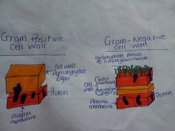
Gram staining is a technique used to see if a bacteria’s cell wall is gram positive or gram negative. Gram-positive cell walls have a thick wall of peptidoglcyan which traps Crystal Violet and makes the cells look a dark purple/maroon color. Gram-negative cell walls only have a thin layer of peptidoglycan which allows the Crystal violet to wash off easily, leaving the cell pink or red. Gram-negative cell walls are more complex because they have an outer membrane that contains lipopolysaccharides. (Carbohydrates bounded to lipids). This lipid portion of the lipopolysaccharide can be toxin, causing fever and shock. Also the extra membrane protects the cell from the body’s defenses which makes it more resistant to
antibodies. This information is very helpful in medicine in that it determines if the persons infection is due to gram-negative or positive bacteria.