Introduction
We performed a two-part experiment to gain a better understanding of bacteria through gram-staining. Gram staining is a differential staining method which results in the classification of two groups of bacteria, Gram-positive and Gram-negative. This technique is based on the fact that the gram-positive bacteria's cell wall has a stronger attraction for crystal violet, based on the presence of more peptidoglycan, then does the gram negative bacteria. We also learned about and applied the aseptic technique to prevent contamination. We viewed E.coli and B. Cereus and determined which were gram positive or gram negative. In the next part of our experiment we swabbed various surfaces, the bottom of our shoes and the classroom light switch. We did so to determine how many and what kind of bacteria could be found there. We transferred these swabs to a petri dish and allowed them to culture for a week. We viewed the petri dishes a week later and determined how many colonies grew, whether they were gram-negative or gram-positive, and the differences in their morphological features.
Background information
Prokaryotes
Bacteria are prokaryotic organisms which lack a nucleus and membrane-bound organelles. Prokaryotes are thought to be organisms living on earth. Most are unicellular, but some species live temperately or permanently in colonies. They are the most widely distributed group of organisms than any other on earth. These tiny organisms are microscopic when looking at them individually. There are three shapes of bacteria, Bacillus (rod-shaped), Coccus (spherical) and Spirillum (Spiral).
Nutritional Mode
Most bacteria are heterotrophs, meaning they cannot make they own food and must get nutrition from organic molecules made by other organisms. It is because of this characteristic that they are called decomposers, and they feed off of dead and decaying matter. There are also Autotroph
bacteria that get their energy from photosynthesis or the oxidation of inorganic
molecules.
Reproduction
Bacteria reproduce asexually when a cell DNA replicates and the cell pinches in half without the nuclear and chromosomal events with mitosis, a process known as binary fission. There is also such thing as bacteria sex. Bacteria can transmit DNA from one other causing diversity.
Pathogenic/Mutualistic Bacteria
Some bacteria are pathogenic, meaning that they cause disease. On the other hand, most bacteria are not. You can find mutualistic bacteria living on and inside of you whom actually help you. For example, your body is covered in bacteria that eat the dead skin and there are also bacteria living
inside of you giving you vitamins.
Hypothesis
For the first part of the experiment we predicted that the E. Coli and B. Cereus bacteria would be gram-negative since they cause food poisoning. From the samples we swabbed, we expected to find two or more different colonies of bacteria. We predicted that at least one would be gram-negative and both would be of different morphology. We predicted that one of the colonies would be irregular and flat while the other colony will be circular and raised.
Procedure One - Aseptic Technique
-Aseptic technique refers to the precautions that should be taken to limit exposure of the worker and the environment to the bacteria. Also it includes the prevention of contamination of the bacteria culture by other organisms.
-In order to properly sterilize the inoculating loop, it should be run under a flame and cooled on a section of the agar before applying to the bacteria quadrant.
-In order to properly sterilize the inoculating loop, it should be run under a flame and cooled on a section of the agar before applying to the bacteria quadrant.
Procedure One - Pure Culture and Transfer Techniques
In the laboratory, transferring a bacteria population of microorganisms to a growth medium (petri dish), is referred to as inoculation. The microorganisms are called the inoculum.
A) We started the inoculation process by labeling our petri dish on the bottom. We labeled the bottom because the tops of the dishes could be switched and the petri dishes are often stored upside down to prevent condensation from forming in the sample. We next rolled the swab of bacteria onto one quadrant of the agar.
B) Next we used a bacterial inoculating loop to spread the inoculating material over the agar to produce isolated colonies. We accomplished this by transferring less culture material into each successive quadrant using gentle pressure.
C) We sterilized the loop by putting it under a flame and cooling it. Then we spread the bacteria from the first quadrant to the second quadrant. After the loop was flamed again the bacteria was spread from quadrant two to three and then from the third to the fourth. We never removed the lid of the petri dish completely so the bacteria was not exposed to the medium of contamination.
Procedure Two - Culturing Bacteria Using Aseptic Techniques
1. We obtained a sterile cotton swab and a closed petri dish containing nutrient agar.
2. We each swabbed different areas, the bottom of our shoes and the light switch.
3. We opened the petri dishes and dragged the swab over the surface of the agar using our procedure one technique.
4. We closed the lid and used perafilm to seal the edges. We labeled the dish with a sharpie and left it for a week.
5. After a week's time we examined the agar for bacterial growth.
Procedure Three - Gram Staining
1. We first marked the spot on the slide where the sample will be placed.
2. We used proper aseptic technique and removed two loops full of organisms from the tube and deposited them on the marked part of the slide.
3. Our culture was heavy so we also put one loop of water onto the slide to dilute the sample.
4. We used thongs to hold the slide and passed it through the bunsen burner to dry and warm it. This step was performed to kill the bacteria and attach it to the slide so it wouldn't wash off during the following steps.
5. We filled three beakers with tap water and arranged the slides on the staining tray .
6. We covered the smear with the crystal violet stain and allowed it to sit for one minute.
7. Next we tipped the excess stain off of the slide and rinsed it by immersing it into each of the three beakers of tap water and moving it around in each.
8. Next we covered the stain with Gram's Iodine solution and allowed it to sit for one minute. We emptied and refilled the beakers of tap water.
9. We removed the excess iodine from the slide and rinsed it in each of the beakers of fresh water.
10. Next we ran ethanol over the slide until the rinsed off ethanol ran clear and let that sit for thirty seconds.
11. We then counterstained the slide with safranin-o solution and let it stand for one minute. We changed the water in the beakers during this time.
12. We then poured off the excess safranin and rinsed the slide in the beakers.
13. Finally we blotted the slide dry, being careful not to rub the smear off of the slide.
14. Finally we were able to view our prepared slide under the light microscope using the oil-immersion technique.
**We repeated this procedure for cultures that we collected by swabbing the inside of our mouths.
Results
Discussion
For the E. Coli and B. Cereus bacteria samples, we found that they were both gram-negative bacteria. This makes sense because we know that gram-negative bacteria can be toxic due to the lipopolysaccharides in their cell wall. This explains the horrible effects of food poisoning that these two bacterium can have on a human. We were not able to view the morphology of these two bacteria.
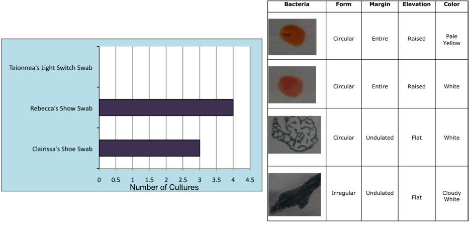
On the other hand, our surface swabs were quite the opposite. We clearly saw the morphology in our samples. In Rebecca’s shoe swab we found four different colonies, in Clairissa’s shoe swab we found 3 colonies which were similar to Rebecca’s, and we did not find any cultures from Teionnea's light switch swab. We were told by our lab instructor that one of Rebecca’s colonies was a fungi. Both Clairissa and Rebecca’s samples contained two different round, flat, and entire shaped bacteria. One was a pale yellow and the other was white. In addition they also had a very large colony that covered the majority of the plate. This bacteria was cloudy white, irregular, flat, and undulated in color and shape. We gram stained three of the colonies and found that they were all gram-positive bacteria.
On the other hand, our surface swabs were quite the opposite. We clearly saw the morphology in our samples. In Rebecca’s shoe swab we found four different colonies, in Clairissa’s shoe swab we found 3 colonies which were similar to Rebecca’s, and we did not find any cultures from Teionnea's light switch swab. We were told by our lab instructor that one of Rebecca’s colonies was a fungi. Both Clairissa and Rebecca’s samples contained two different round, flat, and entire shaped bacteria. One was a pale yellow and the other was white. In addition they also had a very large colony that covered the majority of the plate. This bacteria was cloudy white, irregular, flat, and undulated in color and shape. We gram stained three of the colonies and found that they were all gram-positive bacteria.
Conclusion
In conclusion, our hypothesis were partially correct. We definitely underestimated the amount of bacteria and fungi that would be present on the bottom of our shoes. We gained valuable experience in the process of culturing using petri dishes and gram-staining slides. Some potential error in our experiment was not considering the possibility that the light switch had just been cleaned. This must be the case because we imagine that the light switch would be extremely dirty from regular use. With these results, we have a deeper understanding of how prevalent bacteria are, how they grow, their morphology, and how to determine if they are gram-positive or gram-negative.
